Tuesday, March 31, 2015

Wax Worm/P. larvae LD50

From 3-30-15.

Checked the survival and health of the wax worms injected with and fed the bacterial spores. A number of the wax worms injected with B. thur spores are now dead and displayed large dark sores on their bodies. Images of these sores are shown below. Note: it is much easier to get clear pictures of the wax worms once they are dead!

Dead WW injected with B. thur spores

Dead WW injected with B. thur spores.


//EWW

Wax Worm Maintenance

From 3-24-15.

All of the wax worms in the current generation are either pupating or have already become wax moths. Eggs were recovered and added to a new container filled with wax worm diet that was previously made. There were a lot of eggs that were recovered and the majority were placed inside a 50 mL conical tube and stored at 4C.

There are many more wax worms that will soon emerge as wax moths that will produce many more eggs, so I will keep the eggs at a cold temperature for storage.

//EWW

Sporulation on Blood Agar

From 3-24-15.

P. larvae ATCC #9545 was grown on Colombian sheep's blood agar for seven days at 37C 5% CO2.

1. The vegetative and endospores were extracted from the surface of the agar by adding 5 mL of sterile ddH2O and gently rubbing the surface with a sterile cell spreader.
2. The 5 mLs was collected in a 50 mL conical tube. This wash step was repeated an additional two times.
3. The volumes from three plates were combined into a single 50 mL conical tube. The tube was centrifuged at 18,000 x g for 2 minutes.
P. larvae 9545 on Colombian sheep's blood agar.
4. The supernatant was carefully decanted from the 50 mL tube. The pellet was re-suspended in 3 mL of sterile ddH2O.
5. The 3 mL volume was transferred to 1.5 mL eppendorf tubes in 1 mL volumes. The tubes were centrifuged at at 18,000 x g for 1 minute.
6. The supernatant and upper most layer of the pellet was carefully removed. This uppermost layer is composed of vegetative cells and debris. The endospores are more dense and thus are heavier so are at the bottom of the pellet.
7. The pellet was re-suspended in 1 mL of sterile ddH2O. This wash step was repeated five times.
8. After the last wash and upper pellet removal, the pellet was re-suspended in 1 mL sterile ddH2O again. The suspension was incubated at 60C for 15 minutes in a water bath.
9. The centrifugation and wash steps were repeated an additional five times.
10. QUANTIFYING SPORE STOCK: the suspension was diluted 1:10 down to 10^-8 and 10 uL volumes were plated onto MYPGP agar. Plates are incubated at 37C for 3 days until colonies are counted and CFU/mL is determined.

//EWW


Monday, March 30, 2015

Wax Worm/P. larvae LD50

From 3-27-15.

Checked the survival and health of the wax worms exposed to P. larvae spores.

LD50 Injection

Day 3

Most of the wax worms have begun to pupate and form cocoons. They were late instar larva to begin with, so were likely close to pupating anyways and it is probably not related to the trauma of being injected as the "no inject" controls are also beginning to pupate. There doesn't appear to be any disease in the wax worms that were injected with P. larvae spores, however the wax worms that were injected with B. thur have been showing signs of disease. Namely, most of them have developed black/brown sores on their bodies. A picture of what these sores look like under a dissecting scope is seen below:

Sore on WW body after being injected with 1x10^6 CFU of B. thur spores.
LD50 Ingestion

Day 3

Checked health of the early instar wax worms that were provided spores spiked into 100 uL of BAD. There were many deaths in the B. thur positive control. The P. larvae groups has not had a lot of death yet, however many of the wax worms in this group have begun to display signs of disease. Similar to the sores seen in the late instar wax worms that were injected with spores, the early wax worms have developed unusual colored spots on their bodies. Images of these discolorations are seen below. 

Discoloration of early instar wax worms provided 1x10^4 CFU P. larvae spores
Discoloration of another early instar wax worms provided 1x10^4 CFU P. larvae spores
I will continue to monitor the wax worms survival and health. My supply of this generation of wax worms has begun to dwindle as most are pupating and will soon become wax moths. I am not sure if I will be able to repeat this experiment any time soon..

//EWW

Chlorine Dioxide - Wax Worm Lethality

From 3-27-15.

It has been 48 hours after the late instar wax worms were exposed to the chlorine dioxide gas. The wax worms were allowed to recover from the gas and they were checked for survival today by gentle prodding with a forceps.

Survival Raw Data (n=10):

Replicate A Replicate B Replicate C
0 mg 10 10 10
100 mg 10 10 9
200 mg 7 6 6


Discussion:

The results are similar to the last time I performed this experiment. The most death was seen at the 200 mg concentration, otherwise there was negligible death seen in the 100 mg concentration. It appears the same trend .No wax worms died in the negative control. These wax worms were not exposed to the chlorine dioxide gas. Apparently, soft bodied insects react similarly to chlorine dioxide as certain fruits and vegetables do (ex tomatoes). The bodies of the wax worms serve as a sink for the chlorine dioxide gas as it is high in moisture and provides little physical resistance to absorption.  I am unsure if I will continue to pursue this avenue of experiments.  It would be interesting to see how the gas effects insects with exoskeletons like the red flour beetles or dubia cockroaches we have in the lab...

//EWW

Friday, March 27, 2015

Detection of P. larvae in Local Honey

From 3-21-15.

Attempted to amplify the 16S rRNA gene of the five P. larvae honey isolates and the roach trachea isolate using the 8F and 16Srev (from IDT) primers using a thermocycler parameter I had previously developed based off a paper that used the 8F primer to amply the region. HOWEVER, the gel was completely blank, except for the DNA marker, indicating the amplification was not a success.

I have not been able to successfully amply the 16S using these primers and those parameters in the past, but I believe the problem to be the template DNA. I am confident that the DNA I used as a template is adequate for amplification as it was used successfully previously using the AFB primers. I am now convinced that the issue is either my amalgamated use of primers  or the thermocycler parameters. I will attempt to remedy in the near future as I would like to send the 16S for sequencing soon.

//EWW

Wax Worm/P. larvae LD50

From 3-24-15.

Set up the P. larvae spores LD50 ingestion and inoculation experiment using the early and late instar wax worms larvae again. Unfortunately, I used up the last of my P. larvae spore stock reserves and will need to replenish, hopefully the use of Columbian sheep's blood agar to improve sporulation is successful. I am also running low on the Bee Artificial Diet (BAD) and will have to acquire more from the USDA soon.

LD50 Ingestion

The smallest wax worms (earliest instars) were added to the small containers used previously for this experiment. However, this time only 5 early instar wax worms were added to each container instead of 10. This is in response to the universal decline in health seen in all the wax worms last time due to possible overcrowding and excess frass build up. A volume of 100 uL of BAD spiked with P. larvae spores was still added as before. With the same volume of spores, but reduced amounts of present wax worms, will likely increase the amounts of potential spores ingested by each worm.

Spores added to the container with 5 wax worms:
P. larvae = 10,000 
B. thuringiensis = 1,000,000

100 uL of BAD was distributed throughout the container to avoid large puddle that WW could potentially drown in. By spreading it throughout also increases the likelihood the WW will come into contact with the spores.
B. thur was used as a positive control and was also mixed with BAD when made available to the wax worms. Two negative controls were also implemented, one group was fed only the BAD and another was not provided a diet at all. The containers were sealed with their covers and incubated at 30C in a secondary container. I will check their progress and survival everyday.

LD50 Inoculation 

The larger wax worms were inoculated with 20 uL of P. larvae spores in PBS. Injection spots were near the second to last segment of their body into their hemocoel as done previously. B. thur was used as a positive control. Two negative controls were also used, one set of wax worms were injected with only 20 uL of PBS and another was not injected at all. This will be important to see if making cocoon trends occur between all the groups. Ten wax worms were added to a single petri plate and incubated at 30C in a secondary container. I will continue to monitor their survival in the coming days.

//EWW

Chlorine Dioxide - Wax Worm Lethality

From 3-26-15.

Repeat of the previous ClO2 gas exposure experiment using the late instar wax worms. I exposed 10 wax worms in each container to either no ClO2 gas, 100 mg (total reagent weight), or 200 mg. The reagents were mixed in the modified PCR tubes (six small holes added to them using a heated inoculating needle). The 925 mL volume containers were used again.

Three different conditions:

100 mg 
200 mg

No gas

After the PCR tubes containing the ClO2 reagents were added to the containers they were sealed with their covers and wrapped using Parafilm to ensure the gas could not escape. Containers were incubated at room temperature, protected from light for 24 hours.

I will repeat the procedure I did last time, where I remove the PCR tubes after 24 hours, vent off any remaining gas using the laminar flow hood, and allow the wax worms to recover for an additional 24 hours before determining survival. I will also count the number of wax worms that have formed cocoons (if any) after the initial 24 hours.

//EWW

Tripartite Conjugation in P. larvae

From 3-24-15.

Attempted PCR using the P11/P12 primers and thermocycler parameters described in the previous entry. This is the first time I have used these primers and am using a program that I designed myself. Amplicons were mixed with EZ vision loading dye using two different volumes of DNA since I had been having issues with overloading the wells in the past. Either 5 uL or 10 uL of amplicon DNA was combined with 2 uL of the loading dye and added to the wells of a 1% gel. The gel was ran for 40 minutes at 100 volts. Below is the resulting gel with labels below that:


Lane 1 Lane 2 Lane 3 Lane 4 Lane 5 Lane 6 Lane 7 Lane 8
Hi Lo Transcon pMarA 9545 Transcon pMarA 9545 Empty

Lanes 2 - 4 were the 5 uL volumes of DNA and Lanes 5 - 7 were the 10 uL volumes.

Discussion:

The P11/P12 primers amplify the TnYLB-1 transposon. The "transcon" in Lanes 2 and 5 were a single transconjugate isolate that grew on the MYPGP Poly60 Kan100 plates after the tripartite conjugation. The P. larvae 9545 served as a negative control as it should not contain the transposon that is being amplified. pMarA contains the TnYLB-1 transposon and served as a positive control. The good news is that my thermocycler parameters are more than adequate at amplifying the TnYLB-1 transposon as there were bright and definite bands found in the positve pMarA control. The bad news is there doesn't appear to be a transposon in the transconjugate isolate. This transconjugate isolate tested positive using the AFB primers, indicating that it was P. larvae, however with these results today it means that the conjugation was not successful at inserting the transposon into it.

I may repeat this amplification test using the other transconjugate I have acquired in the future, however it is more likely I will re-attempt the tripartite conjugation and makes changes with the time intervals in hopes of improving my results.

//EWW

Thursday, March 26, 2015

Chlorine Dioxide - Wax Worm Lethality

From 3-25-15.

Checked the survival of the late instar wax worms that were exposed to ClO2 gas for 24 hours and have since been able to recover for an additional 24 hours. Wax worms were checked to be alive or dead through prodding with a forceps. If they moved they were considered alive, if not then they were dead.

Results:

Conc of ClO2 Survival
(n=15)
10 mg 15
100 mg 15
200 mg 3

Survival of late instar WW after exposure to various concentrations of ClO2 gas
Discussion:

All of the wax worms at the lower ClO2 concentration survived the exposure. However, the wax worms at the higher concentration (200 mg) did not all survive. Only 20% survived after only 24 hours of exposure. This is particularly interesting as at half the concentration (100 mg) all the wax worms survived. This study was only a preliminary examination of the effect of ClO2 on late instar wax worms, but it appears I may be close to the concentration at which the wax worms are just becoming susceptible to the gas. If I decide to continue with this experiment, I will repeat this experiment with more replicates.

//EWW

Wednesday, March 25, 2015

Chlorine Dioxide - Wax Worm Lethality

From 3-24-15.

It has been 24 hours since the late instar wax worms were exposed to the chlorine dioxide gas. I did not want to count survival quite yet as I wont be able to tell if the gas did in fact kill the worms or they are just anesthetized. So, I opened each of the containers (removing their lids) and removed the PCR tube containing the ClO2 gas reagents. I also left the open containers in the laminar flow hood for 10 minutes to allow any lingering gas to leave the container. The amount of wax worms that had formed cocoons after just 24 hours of exposure was recorded and listed below. This was interesting because not all the wax worms had done this in every treatment group.

Results:

Conc of ClO2 # of cocoons
10 mg 12
100 mg 12
200 mg 0


Number of WW (n=15) that have formed cocoons after being exposed to ClO2 at different concentrations after 24 hours

The containers were closed once again and stored at room temperature for another 24 hours to allow any of the wax worms that may not be dead to become less lethargic and wake up.

Discussion:

Interestingly, the majority of wax worms exposed to the two lower concentrations of ClO2 reagents seemed to have formed cocoons while the wax worms in the highest concentration. This could be because all the wax worms exposed to the higher concentration are dead and were not able to form cocoons due to time of death. This was very much a preliminary study still, but if I decide to follow up on it I will need to add replicates and more importantly a negative control. I will determine the survival of the WW that were exposed to the ClO2 gas for 24 hours tomorrow.

//EWW


Tuesday, March 24, 2015

Sporulation on Blood Agar

Goal: Improve my ability to extract P. larvae spores and increase their recovered concentration.

Made Columbian sheep's blood agar plates and slants. This type of agar was used by Genersch et al. (2005) for the propagation of P. larvae spores. Their method resulted in a ~10^7 spore concentration. To make the blood agar I autoclaved Columbian agar base and added 5% of defibrinated sheep's blood while stirring after it was cool enough to touch and pour. I made 40 blood agar slants by aseptically transferring 8 mL into sterile glass tubes (picture below). After the hot agar was added to the tubes, the tubes were tilted to achieve the slant. This was done using the racks used in the media kitchen as per Linda P.'s directions.

Columbian Sheep's Blood agar slant
Using a three day old broth culture of P. larvae in BHI, I added 300 uL to the Columbian blood agar slants. An image of what this looks like is seen below:
I will incubate the agar slants at 37C for 10 days, after which I will extract the spores by likely washing the agar slant and recovering the liquid suspension. I also inoculated 10 Columbian blood agar plates with 100 uL of the P. larvae broth culture. The plates are incubating at 37C 5%CO2 for seven days.

//EWW

Wax Worm Maintenance

From 2-21-15.

I made more food for the wax worms to be used in propagation of their next generation. I believe this is about the third or fourth generation of wax worms that I will have that were originally purchased from a pet store. The WW diet ingredients and instructions were designed by Ben M. of the Fisher lab.

Ingredients Needed:
16 oz of Gerber baby food (or equivalent), whole wheat or multigrain
50 g wheat bran or crushed bran flakes
120 mL sterile ddH2O
170 mL Glycerin or Glycerol
100 g Table sugar
800 uL Amphotericin B (to protect food from mold)

Instructions:
1. Heat water and glycerin/glycerol in flask on hot plate using stir bar
2. Add sugar to the hot liquid and continue to spin until all is dissolved
3. Add 800 uL amphotericin B
4. Mix baby food and crushed bran flakes together
5. Add the liquid and stir in quickly
6. Diet should be consistent without lumps.

WaxWorm diet
Prepared diet was stored at room temperature until use.

//EWW


Chlorine Dioxide - Wax Worm Lethality

From 3-17-15.

Performed another ClO2 gas / wax worm exposure. This time using three different concentrations of ClO2 gas. I would like to know the extent to which the wax worms are susceptible to the chlorine dioxide gas, so I will use relatively low concentration this time. Last time, on the 3-17-15 post, I used a relatively high concentration of about 3 grams total weight. Fifteen wax worms were placed into a small container and exposed to a total weight of 10 mg, 100 mg, and 200 mg of chlorine dioxide gas reagents.Below is an image of the size and condition of the wax worms that were used in this experiment:

Fifteen wax worms in the container with a 925 mL volume
The ClO2 reagents were mixed inside of PCR tubes that had been modified with several small holes in them. The holes were made in the PCR tubes by heating up an inoculating needle using a Bunsen burner and passing the needle through the tube. An image of what the tube looked like after the holes were made is below (albeit hard to see):
Holes in PCR tube
A total of six holes were made in each PCR tube (4 on the side and two in the cap). After the reagents were both added to the tube it was briefly shaken and placed inside in the middle of the container hosting the wax worms. The smaller holes in the PCR tube will prevent the wax worms from coming into direct contact with the reagents and still allow the gas to leave the tube and fill the container.

The containers with the wax worms were closed using their lids and sealed using Parafilm. They were stored at room temperature protected from light. I will check the survival of the wax worms after 24 hours of exposure.

//EWW

Tripartite Conjugation in P. larvae

From 1-7-15.
From 3-21-15.

I had previously ordered a couple sets of primers from Integrated DNA Technologies (IDT) on February 10th, 2015. These primers will be used to confirm that the TnYLB-1 transposon is indeed now present in P. larvae after tripartite conjugation. After the primers arrived I resuspended the primers using appropriate volumes of sterile ddH2O and stored them at -20C. I have designed thermocycler programs for each primer set based off a basic temp/time template to start with. The only variable that needed to be adjusted is the annealing temperature, which I have calculated to be within 5 degrees of both of the primers melting temperatures.

pMarA plasmid (from Le Brenton et al.)


Primers:

Primers P11 & P12 were created based on the primers from Xia et al. . These primers will amplify the internal fragment of the TnYLB-1 transposon that is found in the pMarA shuttle plasmid that I am using to transfer the transposon into P. larvae 9545.
P11 (Tm = 56.8C)
P12 (Tm = 56.4C)

Primer oIPCR1 & oIPCR2 were created based on the primers Le Brenton et al..This primer set amplifies outward from the TnYLB-1 transposon.
oIPCR1 (Tm = 45.8C)
oIPCR2 (Tm = 53.0C)

Thermocycler parameters:

Primer sets P11 & P12

Temp Time
Step 1 95.0C 2.00 m
Step 2 95.0C 1.00 m
Step 3 56.6C 1.00 m
Step 4 72.0C 1.00 m
Step 5 GoTo Step 2, repeat x 24
Step 6 72.0C 5.00 m
Step 7 20.0C Forever


Primer sets oIPCR1 & oIPCR2
Temp Time
Step 1 95.0C 2.00 m
Step 2 95.0C 1.00 m
Step 3 49.5C 1.00 m
Step 4 72.0C 1.00 m
Step 5 GoTo Step 2, repeat x 24
Step 6 72.0C 5.00 m
Step 7 20.0C Forever

//EWW

Wax Worm/P. larvae LD50

From 3-21-15.

It has been three days since I injected 20 wax worms with 2000 P. larvae spores (the highest concentration I could get so far), but none of the wax worms have died so far. None even look to be ill or lethargic. Most of them have begun to pupate and form cocoons, which is a phenomenon that I have noticed to occur with all WW injected with anything (even the PBS-only) at about this time after injection.

I will continue to monitor their survival in the coming days.

//EWW

Saturday, March 21, 2015

Detection of P. larvae in Local Honey

From 3-17-15.

Repeated PCR of the honey isolates using the AFB primers. Ran 1% gel, 10 uL amplicon DNA + 2 uL EZ vision dye. Below is an image of the gel ran at 100 volts for 60 minutes.


Lane 1Lane 2Lane 3Lane 4Lane 5Lane 6Lane 7Lane 8
Hi Lo8a2a22a63aQR. Trach9545

The template DNA used in this reaction is the same that was used in the previous PCR using the AFB primers only I used half as much as before. The positive control, P. larvae 9545, amplified using these P. larvae specific primers. The roach trachea (lane 7) served as a negative control as it has not amplified using these primers in the past and is highly suspected to a Bacillus spp. This time with the honey isolates you can clearly see the bands and there is minimal DNA left in the wells (no smearing). All of them amplifed except for 8a (Lane 2), which has amplified in the past indicating that it is indeed P. larvae . However, you can see that there is something in the well of this lane and it could be that I still used too much DNA for that isolate. In the future, I will only use half the amount of DNA that I have been using when setting up my PCR reactions.

//EWW

Wax Worm/P. larvae LD50

From 3-16-15.

LD50 Injection

Day 7 after injection and feeding of P. larvae spores to the late instar wax worms.

Below is the results of the WW injection LD50 study:

3/14/15 3/15/15 3/16/15 3/17/15 3/18/15 3/19/15 3/20/15 3/21/15
Injected: Day 0 Day 1 Day 2 Day 3 Day 4 Day 5 Day 6 Day 7
P. larvae  400 10 10 10 10 10 10 10 9
400 10 10 10 10 10 10 10 10
400 10 10 10 10 10 10 10 10
80 10 10 10 10 10 10 10 10
80 10 10 10 10 10 10 10 10
80 10 10 10 10 10 10 10 10
16 10 10 10 10 10 10 10 10
16 10 10 10 10 10 10 10 10
16 10 10 10 10 10 10 10 10
B. thur 400000 10 0 0 0 0 0 0 0
400000 10 0 0 0 0 0 0 0
400000 10 0 0 0 0 0 0 0
PBS None 10 10 10 10 10 10 10 10
None 10 10 10 10 10 10 10 10

At Day 1: all the positive control Bacillus thur. WW were dead as seen in 3-16-15 post.
At Day 2: I changed the petri plates that all the wax worms were in since there were getting very filthy with frass, the WW were also showing signs of disease (black spots) in the higher spore concentrations
At Day 3: Some have begun to form cocoons
At Day 5: black spots are no longer present in WW afflicted with them earlier
At Day 6: all of the WW are now in cocoons

Discussion:

The positive control WW died right away at Day 1, which is good because it shows that my ability to inject the WW was successful. However, this was an incredibly high concentration of B. thur spores relative to my P. larva spores. It could be related to the shear number of bacterial spores that I injected that caused death and not due to the pathogenesis of the bacteria. But, B. thur is a known insect pathogen. Regardless, the positive control fulfilled its purpose and I will continue to use B. thur as that control for the wax worms.

At day 2 the WW injected with a higher concentration began to show sign of potential disease with the formation of black spots on their bodies similar to what was seen in the early instar wax worms that were fed diet spiked with spores. However, this was about the time the petri plates were covered in copious amounts of frass from all ten of the wax worms and the signs of illness could have been a symptom of their inhospitable environment.

The WW all began to form cocoons as the time progressed. This phenomenon was not seen in the below experiment using the same instar of wax worms except they were fed a spore-spiked diet and not injected. After six days post injection all the wax worms in all the groups (including the PBS negative control) had formed cocoons in the petri plates.

Unfortunately, there was almost no deaths observed in any of the experimental groups (except one in the 1:5 which didn't appear to succumb to a disease). This does not bode well for identifying an LD50 curve. I obviously need to increase the concentration of spores that the WW are injected with, which has been an issue in the past as well. I am working towards an alternative method to culture P. larvae using columbian sheep's blood agar and BHI broth to increase spore yields.

I will re-attempt this experiment in the very near future, but am waiting on WW to grow in size enough to inject. The WW used in this experiment are still in the 30C walk in incubator and I will continue to monitor their survival until they pupate into wax moths in which case they will be disposed of.

LD50 Ingestion

It has been seven days since the late instar WW were fed BAD spiked with P. larvae spores in a volume of 200 uL for 10 WW in a petri plate. The survival was monitored closely for that time. Below is the results of this experiment:


3/14/15 3/15/15 3/16/15 3/17/15 3/18/15 3/19/15 3/20/15 3/21/15
Fed Day 0 Day 1 Day 2 Day 3 Day 4 Day 5 Day 6 Day 7
P. larvae  4000 10 10 10 10 10 10 9 8
4000 10 10 10 10 10 9 9 9
4000 10 10 10 10 10 9 8 8
800 10 10 10 10 10 9 8 8
800 10 10 10 10 10 10 8 8
800 10 10 10 10 10 9 9 8
160 10 10 10 10 10 9 7 6
160 10 10 10 10 10 8 6 6
160 10 10 10 10 10 9 8 7
BAD None 10 10 10 10 10 7 6 5
Only None 10 10 10 10 10 8 7 5
None 10 10 10 10 10 8 8 6

Discussion:

I did not change out the petri plates as I did with the LD50 Injection study above. There was a general trend of death starting at Day 5 that is seen in all the treatment groups, including the negative control BAD only group. There was no difference between the negative control and experimental groups in this experiment. I likely will not repeat the LD50 ingestion using the late instar WW in the future.

//EWW

Tuesday, March 17, 2015

Detection of P. larvae in Local Honey

From 3-11-15.

Re-ran PCR from 3-11-15 using the AFB primers. The lanes are set up as before. 10 ul of ampicon DNA was combined with 2 uL of EZ vision dye and loaded into the well. The 1% gel was ran for 1.5 hours at ~100 volts. The gel is seen below:


Top:
Lane 1Lane 2Lane 3Lane 4Lane 5Lane 6Lane 7Lane 8
Hi Lo8a2a22a63aQR. TrachNone
Bottom:
Lane 1Lane 2Lane 3Lane 4Lane 5Lane 6Lane 7Lane 8
Hi LoTransConjTransConjTransConj
9545
Neg

Discussion:
The good news is that there were nice clean bands seen, the bad news is that there weren't a lot of them (which there definitely should have been). Even the positive control of P. larvae 9545 didn't have a band, which indicates something is clearly amiss. There appears to be a lot of DNA caught in the wells, which may be an indication I am using too much template DNA. Most of the template DNA I used was from a crude genomic prep (heating whole cells), so it is odd that they would result in such a high quantity of DNA. The entire top row was expected to amplify (except for the R. (roach) Trach (trachea) using these AFB primers as they have be shown to in the past. How odd that only a couple of them did. I seem to be having some trouble with PCR lately, it could be attributed to the fact I haven't done it in so long and have lost some of my finesse. I am planning on running more PCR in the near future, but if I am unable to even get my positive control to amplify I am going to have a hard time. So, I will double check my procedure, perhaps I am missing a crucial step or PCR ingredient, or perhaps my thermocycler program has been altered. I will try this applification again using less template DNA, likely diluted 1:10.

//EWW

Chlorine Dioxide - Wax Worm Lethality

From 3-16-15.

Checked on the wax worm health today (~18 hours after ClO2 exposure). Interestingly, the 10 wax worms that were exposed to the chlorine dioxide gas are now all dead and all the ones that were not exposed are still alive and well.

These results confirm my hypothesis, that the cholrine dioxide gas would kill the wax worms. The reasons I am using the gas was to gauge its efficacy at killing P. larvae spores, and possibly use it as a method to treat an American Foulbrood diseased honey bee hive. These results also don't bode well for the likelihood of the gas not killing the honey bee larvae.

All the wax worms were frozen at -20C overnight to kill them and then disposed of in a bio-hazard container.

I may do a dose titration experiment with varying concentrations of ClO2 gas and the wax worms in the future, depending on the quantity of wax worms I have available.

//EWW

Monday, March 16, 2015

Wax Worm/P. larvae LD50

From 3-14-15.

It has been seven days since the early instar wax worms were fed BAD spiked with P. larvae spores. Started on 3-3-15. The raw survival data is saved to an Excel file, but the data is also displayed in the two tables below. Statists were calculated as three replicates of 10 wax worms each.
Raw data results
Normalized data to give percent survival
Concentration of spores that were put onto each plate (taking into account the 100 uL volume)
1:5 = 2000 spores
1:25 = 400 spores
1:125 = 80 spores
The above concentration of spores were added to each container with 10 early instar wax worms in each container. So, it is likely that each wax worm only ingested about one tenth of the concentration above.

Discussion:

Early instar wax worms that were fed the only the BAD without spores had only about 78% survival after seven days, which isn't that bad, but not as good as it should be or I'd like it to be. I believe that the low survival is due to the small storage container that was used. There were 10 wax worms in each small container and the container eventually filled with feces and other debris. Interestingly, the wax worms will eat organic matter, including each other. This is especially evident when there are different sizes of wax worms present in the same container. On one day a wax worm was completely missing from the container when I checked survival. There was no sign of it anywhere and I am absolutely sure it did not escape, however, the container was full of early instar wax worms that were about three times the size of the missing wax worm. I cannot confirm its fate, but I believe it was eaten either to a diseased state making it more vulnerable, or just simply being a runt of a wax worm.

There was a trend of survival seen depending on the amount of spores that the wax worms were fed. The 1:5 dilution resulted in the most deaths and at the quickest rate and so on. However, the error bars indicate that this trait is not statistically significant, meaning there was no distinction between the amount of spores given (1:5 to 1:125) and the survival rate. It is also evident that I should have gone longer beyond seven days to check survival and will likely do so in the future.

I will attempt this experiment again in the future, hopefully with more early instar wax worms and a higher concentration of spores. I need to find a way to categorize the instars of the wax worms and then sort them for the experiment.


LD50 Injection
I checked the late instar wax worms that were injected with P. larvae and B. thur spores yesterday. All of the wax worms that were injected with B. thur are now dead. Pictures of what they looked like only ~18 hours after injection are seen below. They were non-responsive when prodded with a forceps and visibly look dark and deceased. These wax worms were injected with 4x10^5 B. thur spores, which is a very high concentration.

WW injected with Bacillus thuringiensis after one day.

WW injected with Bacillus thuringiensis after one day.
After the above wax worms were disposed of, I realized that maybe they were not dead, but in fact have begun of pupating... It could be that after injection of the B. thur spores triggered pupation in the wax worms. I will most likely repeat this experiment using B. thur as a positive control, albeit at a lower concentration.


//EWW

Chlorine Dioxide - Wax Worm Lethality

The Goal: Determine if chlorine dioxide gas is able to kill adult wax worms (5th instar) at any concentration.

Since I have plenty of wax worms and chlorine dioxide gas components I thought I'd give this a try. This may lead to an effective way to treat wax worm infected hives in the future.

Ten late instar wax worms were placed into two Tupperware containers (volume of 1.2 Liters). One of the containers was not exposed to the chlorine dioxide gas at all, this will serve as a negative control. Below is a picture of the 10 wax worms in the large container.

10 WW in Tupperware container that will be exposed to ClO2 gas
The other container of wax worms will be exposed to chlorine dioxide gas. Approximately 1.5 grams of Part A and Part B chlorine dioxide reagents was added to the small container seen in the picture below. The idea was to keep the wax worms away from coming into direct contact with the reagents and also allow the reagents to mix together. 
Created a container to mix the ClO2 components that the WW will not be able to com into direct contact with. It is wire mesh and a cover. The wire mesh is taped on the back.
The chlorine dioxide gas reagents were added and the above reagent container was added to the center of the 1.2 Liter Tupperware container. The container was covered with its lid and the edges were wrapped with Parafilm. The control and experimental containers were place at room temperature, protected from the light in the Fisher Lab. It is very important that the containers are protected from the light as it can effect the generation of chlorine dioxide gas.

I am fairly certain that the gas will kill the wax worms due to its lethal effect on smaller organisms and mode of killing. I also added a rather large concentration of reagents, so I will be surprised if it doesn't kill the wax worms. But, just to confirm this hypothesis I will test it out.

//EWW

Saturday, March 14, 2015

Wax Worm/P. larvae LD50

From 3-9-15.

Day 5
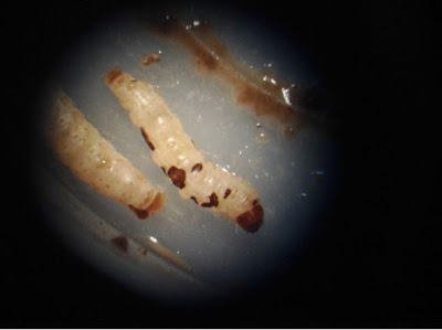
Checked on the early instar wax worms that were fed the P. larvae spores. On day 4 they began to show visible signs of illness on their bodies. This is about the time frame as before. Images of these black/brown sores on their bodies can be seen below. I have also begun to see an increased rate of death in the less dilute spore groups.

Early instar WW with signs of illness (sores on body)

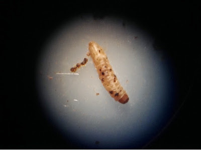
Dead WW that was exposed to the 1:5 P. larvae spore dilution in its diet 5 days ago.

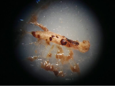
Dead early instar WW. Notice deformity in body were the sores are.
LD50 Injections
I plan on determining the LD50 of P. larvae spores in the later instar WW as well. For the later instars I plan on injecting the spores directly into their hemocoel.


I injected late instar WW with 20 uL of a diluted spore stock using a small needle. I was able to hold the larger WW in place and inject them near the second to last segment of their body (second from anus end).

For a positive control I used B. thuringiensis, a known insect pathogen, and for a negative control I used PBS. For the experimental groups of P. larvae I used three different dilutions (5 fold serial dilution). Below is the amount of spores that was actually injected into each of the wax worm (accounting for dilution and the 20 uL volume that was injected):

Bacillus thuringiensis - 4x10^5 CFU (spores)
P. larvae - 4x10^2 CFU, 8x10^1 CFU, and 16 CFU
Phosphate Buffered Saline (PBS) 1x

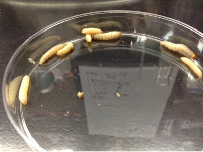
WW after injections, stored in petri plate (10 per plate).
The area were the WW were to be injected were wiped with ethanol and allowed to briefly dry as to avoid any secondary infection (I work in an insect pathology lab!). The injections themselves took place on the lab bench on top of a standard white sheet of printing paper. The paper was switched between treatment groups as to avoid cross contamination that would effect the outcome. 

Comments on injecting WW: Be confident with the needle stab and be sure to insert the needle far enough in that the 20 uL volume doesn't leak out due to a superficial stab. After injecting the volume, let go of the wax worm with the needle still inside of it and then pull out the needle. This prevents putting physical pressure on the WW that would cause the injected volume or hemolymph from coming out of the newly created injection wound.

After injection, the WW were carefully transferred to a petri plate either via careful use of forceps or by rolling. Each treatment group included 30 WW (10 to a petri plate). The WW were placed inside a secondary container and stored in the 30C walk in incubator.


LD50 Diet

Late instar wax worms were also allowed access to Bee Artificial Diet (BAD) that had been spiked with P. larvae spores. No B. thur control was used in this experiment as I am running low on BAD and am not convinced this particular experiment will go anywhere, but we'll see. I am unsure if the late instar WW will even ingest the diet if not force fed and I am also not sure that if they do indeed ingest the spiked diet if the spores will have an affect on their now more functioning immune system. If it does, I also believe it would likely require a higher concentration of spores in their diet to overwhelm their immune system and cause disease. So, the results of this experiment may not tell us anything if the WW all survive, but if they don't then that would encourage definite further investigation. Plus, I had extra BAD spiked with spores from the last experiment and plenty of wax worms to try something with.

Ten WW were sorted into a single petri plate and fed the P. larvae spores in BAD at a volume of 200 uL. The 200 uL were added to the plate in the fashion shown below in the image. This was to prevent drowning of the wax worms in one large puddle of diet as well as to increase the chance that a WW will come into contact with the diet (more spots = better chance)

WW fed 200 uL of P. larvae spore-spiked BAD
P. larvae spores on the plates (accounting for the 200 uL volume actually added):

1:5 dilution of stock = 4000 CFU (spores)
1:25 dilution of stock = 800 CFU
1:125 dilution of stock = 160 CFU

The WW were placed into a secondary container and stored in the 30C walk in fridge.


I will continue to monitor the survival and health of the all the LD50 wax worms for at least a week depending on how survival goes.

//EWW