Monday, May 25, 2015

Wax Worm/P. larvae LD50

From 5-14-15.

I've experimented with a new type of Potato-Honey agar to potentially be used as a media for the P. larvae LD50 assay. The recipe was found on an online blog from some guy who raises wax worms in his basement.

Potato-Honey (PH) Agar (1 Liter)
20 g Agar
20 mL Honey
(Sterilize in Microwave)

A volume of 125 mL of PH agar was made in a 250 mL flask. The solution was microwaved until the potato flakes dissolved and became homogenous (or close to it). The solution was periodically removed from the microwave and stirred by hand. After the PH agar was microwaved a small portion was transferred to the wells of a 96 well plate using a sterile eye dropper. This method seemed to work well and the agar solidified nicely in the wells. My only concern is how sterile the PH agar is and if something will begin to grow in the agar. Remaining PH agar was poured into petri plates, it'd be interesting to see if WW eggs would hatch on this surface...

Side view of 96 well plate with PH agar in the wells
Bottom of 96 well plate with PH agar in it
PH agar is very dense and heavy compared to other agar types (like Nutrient Agar or LB Agar). It isn't as soft and has a rather solid consistency thanks to the presence of the honey. 

//EWW

Wax Worm Maintenance

From 5-5-15.

Wax moth eggs were transferred to new containers with diet. Eggs were present on the tops of the containers near where the lid of the container attaches. A number of the eggs were present on the wax paper at that interface. A hodgepodge of containers were used for new eggs. Most of the wax worms have almost completed pupation and are laying eggs, so I'll continue to monitor and transfer eggs as I see them.


Contamination:
There were two large secondary containers each with four smaller containers within. The containers were stored in the 30C walk in incubator, but the container that was stored in the secondary corner of the room had three of its four smaller containers contaminated with fungus! The other secondary container that was in the middle of the shelf had no fungal growth. Not a big problem with the survival of the colony as there are still PLENTY of eggs in the other containers. In fact, some of the eggs were stored in an Eppendorf tube at 4C.

It would appear that conditions were different enough in the 30C incubator depending on where the containers were that it caused fungal growth. In the future, I will only store the containers in the middle of the shelf just to be safe.

//EWW


Sunday, May 24, 2015

ClO2 generation in chambers

From 5-17-15.

Checked the ppm of Cl that was generated inside the modified anaerobic chambers using a total weight of 100 mg of ClO2 reagent. Hours 3.5 to 6.0 were checked and will be added to what was previously discovered.


Time ppm Cl
0.5 hr 65
1.0 hr 75
1.5 hr 90
2.0 hr 100
2.5 hr 105
3.0 hr 105
3.5 hr 100
4.0 hr 100
4.5 hr 95
5.0 hr 95
5.5 hr 90
6.0 hr 80

As was predicted, based on the previous results, chlorine present in the air inside the chambers peaks and then drops off over the six hours. The peak appears to be around 2.5 - 3.0 hours. The peak ppm of Cl was about 105 and the presence of ClO2 gas inside the chamber is about 0.25 of that (0.25 is about the conversion factor that was determined by David S.). The presence of ClO2 inside the chambers is incredibly low, which it is even more surprising that the spore killing rate is so high. 

Below are the calculations received from the USDA regarding ClO2 gas generation inside the modified anaerobic chambers: 

Assumptions:
Chlorine Dioxide
67.45 g/mol
Temperature
25 C (298.15 K)
Pressure
1 Atm
Total Volume
2.5 Liter
Release Media
10 mg ClO2 per 6 hours at 1 g each of Part A and Part B

Target Concentration
Total
Mass ClO2
Concentration ClO2
Mass of Media
Part A             Part B
μg/L
μg
mg
ppmv
mg
mg
10
25
0.025
3.6
2.5
2.5
50
125
0.125
18.1
12.5
12.5
100
250
0.250
36.3
25.0
25.0
200
500
0.500
72.5
50.0
50.0
250
625
0.625
90.7
62.5
62.5
500
1250
1.25
181.4
125
125
1000
2500
2.5
362.7
250
250
2000
5000
5.0
725.4
500
500
4000
10000
10.0
1450.9
1000
1000
5000
12500
12.5
1813.6
1250
1250

According to our initial predictions, with a total dry weight of 100 mg ClO2 reagents, we expected to see a ClO2 ppmv of 72.5 (note: not ppm of Cl). The amount of ClO2 we're actually generating inside the chambers is way lower than that (<20 ppmv). The reduced concentration is probably attributed to the polystyrene containers that are used along with the presence of the glass slides lining the bottom. These types of materials all serve as sinks for the gas and the above calculations depict the unrealistic ideal conditions.

//EWW

Saturday, May 23, 2015

ClO2 Study - P. larvae Spores on Glass

From 5-15-15.

I was able to count CFU from the MYPGP plates that had the spores that were exposed to ClO2 gas for three hours on them. Spore stock #10 (1.1x10^7 CFU/mL) was initially inoculated onto the glass cover slips. Since a volume of 100 uL was added to the cover slips that means there is a concentration of 1.1x10^6 CFU on them before they were exposed to ClO2.


ClO2 Reagent CFU/mL
0 mg 9.33E+05
50 mg 6.67E+05
100 mg 5.67E+05
200 mg 3.67E+05


There is a decreasing trend seen in spore survival as you increase the concentration of ClO2 reagents. This is a similar trend to what has been seen before after exposing for 6 hours, however the log killing was higher (due to the increase amount of exposure time). The standard deviations aren't very good, likely due to subtle decreases in spores between concentrations. For example, when 200 mg were used to treat spores on glass for 6 hours there was over a 3 log killing rate. It would appear that the length of exposure time has an effect on killing of spores, as well as generation of gas.

//EWW

Sunday, May 17, 2015

ClO2 generation in chambers

Goal: Determine the rate at which ClO2 gas is being generated inside the modified anaerobic chambers. This information will be useful information for my ClO2 exposure study as an effective was of killing P. larva spores on apiary-related surfaces. Based on preliminary results, there appears to be a parabolic trend associated with gas generation inside the chambers, with Cl ppm peaking around 3 hours.

For this study, the modified anaerobic chambers were set up like they would be for the spore-exposure study (50 mL H2O, stir bar, etc.). A total weight of ClO2 reagents of 100 mg (50 mg from each Part A and Part B) was used for these experiments. The reagents were mixed inside a PCR tube and after mixing the tube was placed, open, inside the chambers. The ppm of Cl was checked every 30 minutes and were recorded.


Time ppm Cl
0.5 hr 65
1.0 hr 75
1.5 hr 90
2.0 hr 100
2.5 hr 105
3.0 hr 105


I will continue the experiment with time points after 3.0 hrs up to 6.0 hours. 

//EWW

ClO2 Study - P. larvae Spores on Metal

From metal on 5-13-15.

I am able to count the CFU on the MYPGP from the spores on the metal coupons that were exposed to ClO2 gas for three hours. Unfortunately, I wasn't able to count any colonies from the treatment groups (even from the 10^-0 dilution!) as no colonies grew on the plates.

Spore stock #11 was added to the coupons, which means there was about 1.8x10^5 CFU on the coupon when it was exposed to the ClO2 gas. I was able to count CFU from the spores on metal that were exposed to ClO2 gas, unfortunately it was 2.2x10^4 CFU/mL, which means there was only about a 12% rate of recovery of the spore from the coupons! This was a similar problem that I had with the spores on wood, the spore recovery is very poor. I will need to adjust my re-suspension protocol for these two surfaces (pine chips and metal coupons). Perhaps increasing the time the surfaces are soaked in the 1 mL H2O would help with re-suspension.

Below is an image of one of the MYPGP dilution plates. There were only two colonies on the plate from the 10^-0 dilution. However, duplicate plates did not have any colony growth on them.
Spores on metal exposed to 50 mg weight of ClO2 reagent for 3 hours 
I will repeat this experiment in the future and adjust the re-suspension procedure.

//EWW

Friday, May 15, 2015

ClO2 Study - P. larvae Spores on Glass

From 5-10-15.

Spore stock #10 (1.1x10^6 CFU/mL) was used in a ClO2 exposure experiment for three hours on glass cover slips. A volume of 100 uL was added to sterile glass cover slips as before and they were exposed to ClO2 also as previously done, however, the exposure time was not six hours as before, but only three hours this time.

Three different ClO2 concentrations were used in this three hour exposure experiment: 50 mg, 100 mg, and 200 mg. A control of no ClO2 exposure was also used.


The ppm of chlorine gas in each container was checked using the columns as before by taking a 200 mL volume of air sample out after three hours. Interestingly, the ppm of Cl gas seem to agree with what was observed previously the last time this experiment was ran for only three hours. The ppm were about twice what was seen after six hours. This further indicates that there is likely a parabolic trend with generation of ClO2 gas inside the modified anaerobic chambers.


//EWW

ClO2 Study - P. larvae Spores on Wood

From wood on 5-10-15.
From metal on 5-13-15.

I was able to count colonies of P. larvae that grew on MYPGP agar. These spores were exposed to ClO2 gas on pine chips for six hours.

How to calculate CFU/mL
(CFU * Dilution Factor * Volume Factor)
or
(# CFU) * ( 1 / 10^ dilution factor) * ( 1 / volume plated in mL) = CFU / mL
ex:
3 * ( 1 / 10^ (-4)) *  ( 1 / 0.01mL) = 3x10^6 CFU /mL


ClO2 conc CFU/mL CFU/mL
0 mg 14000 1.40E+04
25 mg 9000 9.00E+03
50 mg 950 9.50E+02
100 mg 200 2.00E+02

Spore stock #14 was added to each pine chip. 1.6x10^6 CFU/mL was the concentration of this spore stock, which means there was 1.6x10^5 CFU added to each pine chip. That also means there was only about a 8.75% spore recovery from the pine chip that wasn't exposed to any ClO2 gas, which is terrible. Ideally, >90% recovery would be preferable. 

I attribute the poor spore recovery in part to the pine chips becoming saturated and portions of the spore stocks soaking through and onto the petri plate where it was drying. Another possible contributor could be due to the properties of the pine chip with retaining the spores once re-suspended. I will have to alter my procedure to account for these possibilities. Either by added smaller volumes of spores at a time and allow them to dry before adding the entire volume. I may also try to locate some thicker pine chips to use that wont become saturated as quickly with only 100 uL volume.


Also, there appeared to be a lot of different colony morphologies present on the MYPGP agar plates, more than usual for P. larvae. It could be related to other endospores already present in the pine chips that survived the autoclave sterilization process.

//EWW

Thursday, May 14, 2015

Wax Worm/P. larvae LD50

From 5-8-15.

I added wax to three 96 well plates. Bee's wax was cut to small shavings using a sterile scalpel in a petri dish. Every pre-caution was taken to not contaminate the wax beyond its natural state. The wax itself is not sterile, but I didn't wish to add to it! The wax shavings were transferred to the 96 well plates using a sterile forceps.

Only the first and last four columns of each plate had wax added to them. This is because when the wax worms will be added (and an adhesive cover added) it will be easier to access them if they are not in the middle of the plate. The plates were heated in a ~65C water bath for ~5 minutes or however long it took to melt the wax to the bottom of the plate.

Hot plate water bath used to melt the wax in the 96 well plates. Notice, only the first and last 4 columns have was in them.

Bottom of 96 well plate after wax had melted. Only enough wax was added to cover the bottom of the well. In this case, less wax was preferable to more.
The plates were stored at room temperature. The spores have yet to be added to the wax, along with the early instar wax worms.

//EWW

Wednesday, May 13, 2015

ClO2 Study - P. larvae Spores on Metal

Spores on glass from 5-10-15.

Beekeepers use a number of metal tools when maintaining their hives (crowbars, scrapers, shovels, spades, smoke canisters, etc). The efficacy of ClO2 gas as a disinfectant of P. larvae spores on metal will be determined. I acquired 500 cut stainless steel coupons from NDSU shop foreman Kyle R. The coupons are made of 301 grade steel cut to 0.5 inch squares. P. larvae 9545 spores will be used in this experiment initially, followed by the North Dakota isolates.

100 of the metal coupons were autoclaved using the dry cycle in order to sterilize them. Spore stock 11 (1.8x10^6 CFU/mL)was used for this experiment. A 100 uL volume of spore stock was added to the tops of the sterile metal coupons. The spores were allowed to adhere to the surface of coupons by incubating at room temperature for 1 hour in a biological safety cabinet. 

100 uL of spores on metal coupons
The experiment was set up as before, with three coupons being placed inside a single modified anaerobic chamber. Total dry weights of chlorine dioxide reagents were mixed in PCR tubes and the reaction was allowed to take place for three hours this time. This is a change from the previous experiments that ran six hours.

After three hours, the concentration of chlorine gas was determined using the columns once again. 

Chlorine concentration in each container after 3 hours. The numbers indicate what the total weight of reagents were (in mg)
Dry wt Conc Cl
0 mg 0
50 mg 50 ppm
100 mg 100 ppm

The detected ppm of chlorine was very surprising when compared to what has previously been seen using the glass cover slips at 6 hours. The 50 mg concentration of chlorine was pretty close to what had been previously seen, but the 100 mg concentration resulted in twice the detected ppm of chlorine gas after only three hours compared to the previously performed six hours. It could be that the gas peaks earlier than six hours and then lows due to exposure to the 50 mL of H2O being present. I could conduct an experiment of the gas generation inside the chamber in the future, but for now I will just continue with these experiments and see if this new trend continues.

The metal coupons treated with ClO2 gas were re-suspended in 1 mL of sterile ddH2O. The coupons were allowed to incubate at room temperature in the water for 5 minutes before diluted in H2O and plated on MYPGP agar. The plates were incubated at 37C for four days before colonies can be counted.

//EWW

Sunday, May 10, 2015

ClO2 Study - P. larvae Spores on Wood

From glass on 5-8-15.

Performed ClO2 exposure to P. larvae 9545 on pine wood chips.

A 100 uL volume of Spore Stock #14 was added to the top of a small pine chip purchased from Tractor Supply. The volume was allowed to evaporate and adhere to the chips (placed in petri plates) in the biological safety cabinet for one hour. The volume was rather large for the size of most of the chips. After the one hour time, some spots could be seen under the chips on the petri plate. It appears the 100 uL volume was enough to fully saturate the pine chip and some soaked through to the bottom of the petri plate. I hope I did not lose too much of the spores on the plates.


Three pine chips with adhered spores were transferred to modified anaerobic chambers. A volume of 50 mL of ddH2O was also added to the chambers to increase humidity. Dry weight concentrations of ClO2 reagents were mixed in PCR tubes and place within the chambers.

Total dry weights of reagents:
0 mg
25 mg
50 mg
100 mg

After the reagents were combined in the PCR tubes and mixed the tube was placed within the chamber (while open) and the chamber was sealed using the cover. The metal stir bar was used again inside the chambers in order to mix the gas more effectively. The chambers were protected from light and incubated at room temperature for six hours.

Unfortunately, the magnetic stir bar was not longer spinning in the 100 mg container after six hours. The bar apparently spun out of control and knocked over the petri plate containing the pine chips and the PCR tube containing the reagents. This prevented a good mix of the two reagents, and likely I will not be able to use the results from this treatment group. The ppm of chlorine gas was checked in each container after six hours.

ppm of Cl in each container after six hours:
mgppm Cl
25
15
50
20

Temperature inside the hood: 28.3 C

The determined ppm inside the containers were about half of what they were when using the glass cover slips. The reduced ppm of Cl in the containers could be due to the presence of the pine chips. It could be that they are serving as a sink for the gas similar to how water does.

The pine chips were removed from the modified anaerobic chambers and re-suspended in 1 mL of sterile ddH2O in 50 mL conical tubes. The tubes were vortexed for 30 seconds and left to incubate at room temperature for 5 minutes. The volume was diluted and plated onto MYPGP agar. After about five days I will be able to count colonies and determine CFU/mL of the spores subjected to ClO2 treatment.

ND Isolate Spore Stocks

The P. larvae spores extracted from ND isolated (from honey) were determined today once the colonies were countable on the MYPGP agar. Below are the calculated CFU/mL concentration of these spores stocks. The stocks are stored in 4C and will be used in the near future for the ClO2 study.


ND Stock # CFU/mL
2a -A 1.56E+06
2a -B 1.03E+06
4a -A 7.30E+04
4a -B 3.50E+04
5a -A 3.25E+06
5a -B 2.25E+06
22a -A 5.00E+06
22a -B 3.75E+06
55a-A 7.50E+05
55a-B 7.75E+05

//EWW

Friday, May 8, 2015

Wax Worm/P. larvae LD50

From 5-6-15.

While the wax worms are continuing to pupate into wax moths and lay eggs I am continuing my investigation into how to use bee's wax in place of the oats in the LD50 experiment.

Since the 96 well polystyrene plates with the wax in them would need to be heated to too high of a temperature in order to melt the wax I am moving on to other methods.

My initial plan was to melt the bee's wax inside of a larger container and then quickly pipet the liquid wax to the wells. A small block of bee's wax was placed inside an Eppendorf tube and PCR tube (seen below) and heated in a water bath at 60C for 1 hour. However, at this temperature the wax did not melt and still appeared as the first picture below. By increasing the temperature to 70-80C the wax melted in almost 30 seconds after being placed inside the water. The melted wax inside the tubes is also seen below.

Solid bee's wax inside Eppendorf and PCR tubes
Melted bee's wax after incubating at 70-80C for 30 seconds in a water bath
Unfortunately, I was not able to pipet the wax as it cooled almost instantly as soon it was sucked up into the pipet (really neat!). I was not able to eject the wax into the wells since it was solidified and stuck inside the pipet. I could possibly try heating the pipets themselves, but instead I moved on to another method.

Small slabs of bee's wax was cut using a razor blade as seen below and transferred to a well in a 96 well polypropylene PCR plate (also seen below).
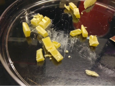
Cut bee's wax to be put into 96 well plate

96 well polypropylene PCR plate
The 96 well plate was placed inside the same plate heater previously used with the polystyrene plate, except this time water was added inside the plate holder to help equalize the heating process. At 60C the wax did not melt, but when pushed to ~70C the wax melted fairly quickly to the bottom of the well. This process seemed to work very well, except I will need to do more with standardizing the amount of wax that is added to each well as some have visibly more than others due to the amount initially added.

There were some air bubbles below the wax in some of the wells when the wax wasn't initially pushed all the way to the bottom of the well. Also, as soon as the plate of melted wax was removed from the water bath the wax solidified (almost) instantly. The wax was still soft enough to easily poke a hole through it with a toothpick. Now that I have a method to melt the wax, I need to determine a way to incorporate the spores without destroying them in the process (due to heat).

//EWW