Saturday, January 31, 2015

Detection of P. larvae in Local Honey

From 1-24-15.

Crude genomic lysate was prepared according the Crude Genomic Lysate for PCR protocol of the dd1-8a, dd5-Q, dd8-63a, dd11-2a,dd12-22a, and the roach trachea isolates. Primers 8F and 1492R (16S rRNA) were used in a PCR reaction using the lysate as DNA template. Amplicons were ran on a 1% gel (seen below). 10 uL of amplicon + 2 uL EZ Vision loading dye. Hi Lo DNA marker was used.


Lane1 Lane2 Lane3 Lane4 Lane5 Lane6 Lane7 Lane8
Hi Lo dd8-63a Roach dd5-Q dd11-2a dd12-22a dd1-8a H2O

Discussion:

It turns out that the issue I have been having with blank gels can be linked to an issue with the DNA template since there were amplicons on the gel that was ran above. Ampliconsc an be observed in lanes 3 - 6, but not in lanes 1 and 7. The Hi Lo DNA marker isn't very clear, which is odd since I can usually determine fairly easily where the bands are located. It appears that the amplicons appeared around the 1500 bp marker, not unusual for a 16S rRNA gene. I am not certain why no bands appeared in Lanes 1 and 7. Perhaps there was an issue with the crude genomic lysate that was used, maybe there wasn't enough template, since there should have been amplification.

In conclusion, it appears that the issue I was having was with the genomic DNA isolation kits, and that the primers and PCR parameters are functioning appropriately. I will next use a different DNA isolation kit to extract genomic DNA and then run PCR.

//EWW

Chlorine Dioxide Pilot Study

From 1-24-15.

Counted CFU on P. larvae spore plates:


Spore Stock 10^-2
D 1 3 4 1 2
E 3 1 3 2 1
F 0 1 1 0 1
G 5 8 3 5 6
H 7 5 10 9 11
I 3 4 2 3 4
J 2 1 1 0 1

Spore Stock 10^-1
C 2 3 2 1 2
Equation used to calculate concentration of Spore Stocks:
[AVG # colonies counted / volume pipetted onto plate in uL] * [ 1 / serial dilution made (ie 10^-2)] * [1000 uL / 1 mL] = CFU/mL

Concentration
Stock C = 2,000 CFU/mL
Stock D = 55,000 CFU/mL
Stock E = 20,000 CFU/mL
Stock F = 6,000 CFU/mL
Stock G = 54,000 CFU/mL
Stock H = 84,000 CFU/mL
Stock I = 32,000 CFU/mL
Stock J = 10,000 CFU/mL
(Stocks are in a 500 uL volume of H2O)

Followed procedure on 10-28-14.

Goal: Determine if chlorine dioxide gas is able to kill P. larvae spores at any concatenation. For this study, a high and low concentration of chlorine dioxide gas will be exposed to spores on glass cover slips for a period of time. After exposure, CFU will be determined via plate counts. This is an important preliminary step to determine the efficacy of chlorine dioxide gas as a sterilization agent of P. larvae spores.

Re-concentrated spore stocks H and I by pelleting them and re-suspending at 200 uL H2O. Sterile glass cover slips were inoculated with 50 uL of spore stocks (volume was placed on the center of the square glass cover slip) and were allowed to dry for about an hour in the fume hood.

Reagents for Chlorine Dioxide gas, Part A and Part B as seen below, were weighed out using the balance in the Pruess Lab. Two different concentrations were weighed out: 50 mg and 5 mg of each reagent. The reagents will be mixed 1:1 to generate the gas in a 50 mL conical tube. For example, 50 mg of Part A will be mixed with 50 mg of Part B, with a final concentration of 100 mg of reagent.
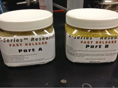
ClO2 reagents
After the two chlorine dioxide reagents were combined in the 50 mL conical tubes the glass cover slips containing the dried spores were quickly and aseptically transferred to 50 mL tubes (image below). Note: be careful that the ClO2 regents are at the bottom of the tube to ensure better interactions with each other and to generate gas more efficiently.

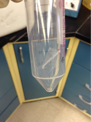
Glass cover slip in 50 mL conical tube
There were triplicate tubes containing the 100 mg reagents, 10 mg reagents, and no reagent at all (control). I fear that even the 10 mg reagent mix might still generate gas at too high a concentration that I wont be able to quantify any surviving spores as they will all be killed. Not necessarily a bad thing, as it shows just how sensitive even low concentrations of ClO2 are to the gas, but it makes my survival curve difficult! Also, I still need to more accurately calculate the ppm of chlorine dioxide gas in this volume of tube and reagent amount.

The tubes were sealed with parafilm and transferred to a shaker incubator protected from light. Temperature was set to room temp (not on) and tubes were gently shaken for six hours. It is important that the tubes are protected from direct light as much as possible once the reagents were mixed as the light can dissociate the gas. It is also important to rotate the tubes in an attempt to better mix the gas.

After six hours, the glass cover slips were aseptically transferred to a new 50 mL tube containing 1 mL of ddH2O. The idea is to re-suspend the spores present on the glass cover slip in the H2O. The glass cover slips were broken with forceps each time and vortexed for 30 seconds. Tubes incubated at room temperature for 5 minutes. 10 uL volumes of the tube culture was plated onto MYPGP agar for CFU counts. Cultures were also diluted 1:2 and 10 uL volumes also plated.

Plates were incubated at 37C for three days until colonies are visible and are able to be counted.

//EWW

Saturday, January 24, 2015

Detection of P. larvae in Local Honey

From 1-22-15.

Ran 1% gel with AFB amplicons from the gDNA and the gel was blank again. So, enough messing around with this gDNA as it is apparently a poor template and I likely made a mistake during the genomic DNA extraction process. I am uncertain what step a mistake was made at. It was likely during the pre-treatment of Gram positive bacteria that was performed. I will double check the method again and repeat it with more stringency in the near future.

I have already struck out the cultures from freezer stocks and am waiting for them to grow at 37C.

//EWW

Chlorine Dioxide Pilot Study

From 1-22-15.

Re-plated the spore stock dilutions onto new MYPGP agar plates and incubated them at 37C. I need to quantify the spore stocks before I can use them in a pilot study.


//EWW

Thursday, January 22, 2015

Detection of P. larvae in Local Honey

From 1-19-15.

Double checked the primers 8F and 1492R and they were correct and so was the PCR parameters from Turner et al. 1999. These parameters were said to have resulted in amplification of the 16S using the 8F and 1492R primers that are also described in the Turner paper. So, it would appear that the primers themselves nor the thermocycler parameters were to blame for the blank gel.

I re-ran the amplicons, loading twice the amount of DNA and also adding EZ vision loading dye, however it still resulted in a blank gel, meaning that the DNA did not amplify. This is possibly because there wasn't enough or any DNA template for the reaction to occur.


//EWW

Chlorine Dioxide Pilot Study

From 1-18-15.

The MYPGP plates incubating at 37C have dried out remarkably quickly. These plates had the dilution series of the spores stocks plated on them to determine the CFU/mL of each of the stocks. The plates are so dried out to the point of almost being crispy, making colony counts difficult and unreliable.

It did seem very dry inside the incubator, with other lab peer's plates visibly dry as well. The plates were not any thinner than other plates I had previously used. However, they were a couple months old... They were only in the incubator for three days, but in the past I have been able to incubate the plates for over a week and the plates didn't dry out. Below are a couple pictures of what the plates and colonies look like:


I will re-plate the dilutions once I make fresh MYPGP plates.

//EWW

Monday, January 19, 2015

Detection of P. larvae in Local Honey

From 1-18-15.

Ran PCR amplicons on the 1% gel (100V for 40 minutes). However, it was a blank gel with only the Hi-Lo DNA marker showing up. None of my amplicons were present on the gel. There are a number of different possible reasons for this.

I only loaded 5 uL of amplicon DNA in the gel and it could be that that volume just wasn't enough to be visualized on a gel. I also did not mix the amplicons with EZ vision loading dye as I was certain that the 2x PCR supermix that was used already contained loading dye. I could try to add the EZ vision dye to a larger volume of amplicon DNA and re-run the gel to determine if either of these factors were responsible for my blank gel.

Additionally, it could simply be that the PCR program that I created in the thermocycler based on what I found through literature review using the same primers was entered incorrectly, or that I mistakenly ordered the incorrect primers (or made a mistake with the primer sequence). This would possibly cause amplification to not occur, resulting in no amplicon bands on a gel. I will double check the primer sequence as well as the thermocycler program just to be sure that those factors were not responsible for my blank gel.

It could be that I made a mistake with the initial genomic DNA isolation. Perhaps I accidentally skipped a step, or used a wrong reagent. I will attempt to quantify the DNA present in my gDNA stocks, something that I should have done initially any ways. I could try to use this gDNA for a different PCR amplification using different primers of known success just to determine if the DNA is viable for amplification. I did originally use a crude genomic lysate of each of these isolates and the AFB primers (16s of P. larvae) successfully before.

//EWW

Sunday, January 18, 2015

Detection of P. larvae in Local Honey

From 1-12-15.

Performed PCR on the six isolates: dd1-8a, dd5-Q, dd8-63a, dd11-2a,dd12-22a, and the roach trachea isolated using the primers 8F and 1492R and the thermocycler parameters previously described. 

Created a 1% agarose gel (0.5 g + 50 mL TAE buffer) and stored it at 4C until use tomorrow.

//EWW

Chlorine Dioxide Pilot Study

From 11-9-14.

Gear up to perform another pilot study to determine if chlorine dioxide gas has any effect at all on killing P. larvae spores.

First I need to quantify the concentration (cfu/ml) of P. larvae spores that I have in my previously created spore stocks. Spore stocks C, D, E and spore stock created on 11-4-14 (called F, G, H, I, J) were diluted 1:10 in a 96 well plate (10 uL  --> 90 uL) down to 10^-4. A volume of 10 uL from each dilution was plated onto MYPGP agar and incubated at 37C.

//EWW

Monday, January 12, 2015

Detection of P. larvae in Local Honey

From 1-11-15.

Created a thermocycler program for the new primers 8F and 1492R based on parameters described by Turner et. al 1999.

The program was named "8F" after the forward primer. The parameters are as follows:

1. 95C - 3 minutes
2. 95C - 15 seconds
3. 49C - 15 seconds
4. 42C - 90 seconds
5. Repeat 2-4 35x
6. 72C - 5 minutes
7. 20C - forever

//EWW

Sunday, January 11, 2015

Detection of P. larvae in Local Honey

From 1-10-15.

Genomic DNA (gDNA) was extracted from the six brothed cultures by using the QIAGEN DNeasy Blood & Tissue Kit. The protocol for pretreatment of Gram-Positive Bacteria was used (enzymatic lysis buffer was previous prepared and used from 10-17-14). The heat block was used for the 37C and 56C incubation steps. gDNA was frozen at -20C until use.

Received primers that were ordered from Integrated DNA Technology recently. The primers are named 8F and 1492R (Turner et al. 1999). These are universal 16S primers that are commonly used for sequencing.

8F
5'-agagtttgatcctggctcag-3'
Tm = 54.3C
MW = 6,148.0
23.7 nm

1492R
5'-ggttaccttacgactt-3'
Tm = 49.4C
MW = 5,784.8
26.7 nm

I resuspended each primer in DNase-free ddH2O according to their nm. Primer 8F was resuspended in 237 uL (23.7 nm) and primer 1492R was resuspended in 267 uL (26.7 nm). Primers were very gently and brielfy mixed by hand in order to ensure thorough suspension. Primers were stored at -20C in my primer box until use.

//EWW

Saturday, January 10, 2015

Detection of P. larvae in Local Honey

From 1-8-15.

It took two days before isolated colonies could be visualized on the plates, incubating at 37C. That isn't odd considering they are highly speculated to be P. larvae and that is about the time it takes P. larvae ATCC 9545 to have visible colonies on MYPGP agar.

Inoculated a single colony from each plate into 10 mL of BHI broth. Incubated at 37C 225 rpm. I need a broth culture of the bacteria in order to extract genomic DNA (no more than 10^9 CFU/mL).

Below are a couple images of what the plates looked when an isolated colony was removed:


Five of the isolates that were brothed were representatives from different honey sources. The sixth isolate that was brothed was isolated from the trachea of a cockroach by Laura N. It is speculated to be Bacillus spp or a yeast spp.

//EWW

Thursday, January 8, 2015

Detection of P. larvae in Local Honey

From 1-5-15.

There was visible growth in all the remaining honey isolates except for isolate number 12, which still had that mysterious globule formations (see previous post). Freezer stocks were temporarily stored at -20C for the time being.

The only isolate from the original honey bee samples that I was not able to make freezer stocks from was isolate number 12 (dd8-85a). However, isolate number 11 was also dd8-85a and was most likely a clone of the isolate 12.

Made isolation streak plates of representative P. larvae isolates from five of local honey samples. There were only five different honey sources that P. larvae isolates were identified from. Isolates were struck out onto MYPGP agar and incubated at 37C. Those isolates were:

dd1-8a
dd5-Q
dd8-63a
dd11-2a
dd12-22a

I plan to extract genomic DNA of these isolates and then sequence their 16S DNA to confirm their identity.

//EWW

Wednesday, January 7, 2015

Tripartite Conjugation in P. larvae

From 1-4-15.

Checked plates that had tripartite conjugation. A number of the plate had colony growth on them!
Each of these plates were incubated at 37C on MYPGP Poly60Kan50 for three days, however growth was only observed on plates that were conjugated previously at 37C. Below is a table illustrating the amount of plates that had a number of colonies on them after the three day incubation.


Temp Disc Broth
RT 0/3 0/3
30C 0/3 0/3
37C 3/3 3/3

Below are images of three of the plates that had a number of colonies on them.


Discussion

It would appear that the tripartite conjugation was a success. That we are able to conjugate the transposon of pMarA into P. larvae 9545 using the pRK2013 helper plasmid. I will determine if the present colonies are indeed P. larvae using the diagnostic AFB primers and I would also like to determine if the original pMarA plasmid is no longer present in its entirety and that its transposon has been inserted into the P. larvae genome.

//EWW

Monday, January 5, 2015

Detection of P. larvae in Local Honey

The From 1-3-15.

Checked the growth of the re-inoculated isolates in the 37C 225 rpm shaker incubator. Most still did not have visible growth. Only isolate 09 had visible growth. Freezer stocks were made of this culture. All others were continued to incubate. I will continue to monitor their progress.

Interestingly, visible clumpy masses were observed in isolate 12. I have no idea what these clumps are, but below are two images of the same tube showing the three masses. The two bottom masses are almost perfect spheres or globules that moved in the growth when shaken. The mass on the surface looks like a small piece of folded paper made of bacteria. This tube was clear yesterday.


This was a really neat observation, but likely the tube will simply be discarded.

//EWW

Sunday, January 4, 2015

Tripartite Conjugation in P. larvae

From 1-3-15.

The 1:1:1 mixture tubes and inoculated filter discs had been incubating at three different temperatures overnight. 100 uL spread plates were made of each broth mixture onto MYPGP Poly60 Kan50 plates. Plates were inverted and incubated at 37C. It may take a few days before visible colonies are observed.

The inoculated discs were aseptically removed from the agar plates using sterile forceps and transferred to 5 mL of ddH2O. Tubes were vortexed briefly to dislodge bacteria from the disc and 100 uL spread plates were made of the suspension on MYPGP Poly60 Kan50. Plates were also inverted and incubated at 37C.


pMarA plasmid from Le Brenton 2006.


Interestingly, the P. larvae 9545 that was inoculated into BHI Poly60 two days ago and that has since been incubating at 37C 225rpm now has visible growth. Yesterday there was no growth that could be seen with the naked eye, however P. larvae grown in BHI only did have visible growth. It appears that the antibiotic slows the growth of P. larvae.

Growth of P. larva 9545 in BHI Poly60

//EWW

Saturday, January 3, 2015

Tripartite Conjugation in P. larvae

From 1-2-15.

Checked broth cultures in the 37C shaker incubator. pRK2013 and pMarA grew to a visible turbidity. However, the P. larvae 9545 culture inoculated in BHI Poly 60 did not have visible growth. This is contrary to what has been determined previously, that P. larvae 9545 is resistant to polymyxin antibiotic. The P. larvae inoculated into BHI only broth did have visible growth. Below is the comparison image of both P. larvae cultures. Both cultures were inoculated from a single colony of P. larvae on MYPGP only agar and incubated at 37C 225rpm overnight.
P. larvae 9545 in BHI broth. Left is BHI only, Right is BHI Poly 60
The P. larvae grown in BHI only was used for this experiment along with the pRK2013 and pMarA cultures. The P. larvae culture grown in BHI poly 60 ug/ml was continued to incubate at 37C 225rpm to see if the culture simply needed more time to have visible growth.

Setting up liquid tripartite conjugation:

1600 ul of each overnight culture was transferred to a 2 ml microfuge tube and centrifuged at room temperature, 13,000 rpm, for 1.5 minutes to pellet. Supernatant was discarded and the pellet was re-suspended in 1000 ul of ddH2O. Pellet was washed three times to remove any excess antibiotics from the original broth media.

The final pellet was re-suspended in 1000 ul of BHI only broth. A 1:1:1 mixture (200 ul of each) was made using each of the three culture; P. larvae, pMarA, and pRK2013. The mixtures were made in a 1.5 microfuge tube. Triplicate tubes were incubated at room temperature, 30C, and 37C. These tubes were NOT shaking and will remain incubating overnight.

Setting up solid tripartite conjugation:

The solid conjugation makes use of  the 1:1:1 mixture that has been previously made. Sterile filter discs were aseptically transferred to MYPGP agar. A volume of 50 ul of the mixture was inoculated onto the disc on the agar. Triplicate plates were incubated at room temperature, 30C, and 37C overnight. Plates were not inverted.

Inoculate 50 uL of mixed culture to filter disc
Aseptically transfer sterile filter disc to agar

//EWW

Detection of P. larvae in Local Honey

From 1-2-15.

All of the remaining cultures that were left at 37C 225rpm to grow still did not have visible growth.
List of isolates that did not grow:
EW code:
01
03
04
05
09
11
12
13

So, the cultures were re-inoculated. Unfortunately, not many (almost none) visible colonies remained on the old plates stored at 4C. So, 5 mL of BHI broth was flooded onto the agar plates and the top of the agar was agitated using a sterile cell spreader (image below).


The inoculated broth cultures were transferred to a 15 mL tube and incubated at 37C 225 rpm. It will likely take a couple days before visible growth is observed. I will make freezer stocks of these cultures tomorrow.

//EWW

The Waxworm & the P. larvae

From 12-24-14.

10 days after transfer:
Checked on the wax worm containers stored in the 30C walk in incubator. They appear to be doing well- no mold formation or condensation. The food appears to be dry and wax worms are lively. There was only one container where the wax worms have begun to pupate and form cocoons.


There is still remaining food in each of the containers and it appears that the wax worms are of a size to begin pupation soon. Interestingly, some of the wax worms are the largest that I have ever seen before. I will continue to monitor their progress. For now they remain in the 30C walk in incubator.

//EWW

Friday, January 2, 2015

Tripartite Conjugation in P. larvae

From 12-27-14.

Broth cultures were made of P. larvae 9545, pMarA, and pRK2013 in 10 of BHI broth with appropriate antibiotics.

9545- Poly60
pMarA- Kan50 + Amp100
pRK2013- Kan50

Cultures were incubated at 37C 225rpm.

//EWW

Detection of P. larvae in Local Honey

From 12-24-14.

Goal: Make freezer stocks of the new P. larvae isolates from local honey.

12-31-14 Wednesday
Inoculated each of the 30 PCR-confirmed P. larvae isolates into 10 mL of BHI broth and incubated at 37C 225 rpm.

1-1-15 Thursday
Most of the cultures had not grown yet. The media was still a clear color. This may be due to the fact that the plates that the isolates were transferred from had been in the 4C fridge for over a week. The cultures may just need more time to incubate, as they are relatively slow growing bacteria. The cultures continue to incubate at 37C 225 rpm.

1-2-15 Friday
The majority of the cultures had visible growth. Freezer stocks were made of these cultures

Freezer stocks: 200 uL 80% glycerine + 800 uL broth culture

The other cultures that had no visible growth were continued to incubate at 37C 225rpm. If no visible growth is observed tomorrow, I will re-inoculate the culture from the plates stored at 4C.

//EWW